Your OWN Dentist
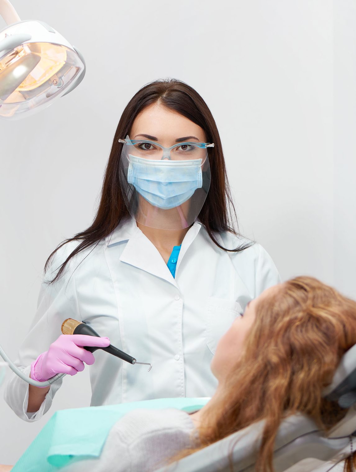
Meet Dr. Rajkumar, a highly skilled and experienced dentist based in the Goregaon suburbs of Mumbai. With 22 years of experience in the field, Dr. Rajkumar has earned a reputation as one of the best dentists in the city.
Our USPs - Quality Assurance Single Sitting Root Canal Fast Treatment Latest Tech

Quality Assurance
Single Sitting Root Canal

Fast Treatment

Latest Tech
Dental Treatments
What Service We Offer
Baghels Dentalworld is efficient in providing comphrensive dental care in the most in the most efficient way following all the necessary protocols.
Dental Anxiety
Dental anxiety is the dental fear or anxiety one feels about visiting the dentist for and conducting a dental treatment.
Dentures
Dentures are removable replacements for missing teeth. They are designed with an acrylic base to fit over the ridges.
Full Mouth Rehabilitation
Full mouth rehabilitation is a step-by-step approach to designed to deliver a beautiful smile with a harmonious bite.
General Dentistry
A general dentist is your primary dental care provider. They treat you for a wide range of dental health problems.
Gum Care
Gum disease refers to inflammation and infection of the tissues surrounding the teeth.
Oral Surgery
Oral surgery includes procedures performed to treat the problems inside the mouth or jaw.
Root Canal
The root canal is a part of the tooth that cannot be seen from the outside, it contains nerves and blood vessels.
Teeth Whitening
It is a simple and effective way to get pearly white smile. It’s a quick and affordable cosmetic dentistry treatment.
Teeth Implants
Veneers are thin coverings that are placed on the front surface of the teeth to improve the teeth’s appearance.

Best Of The Best
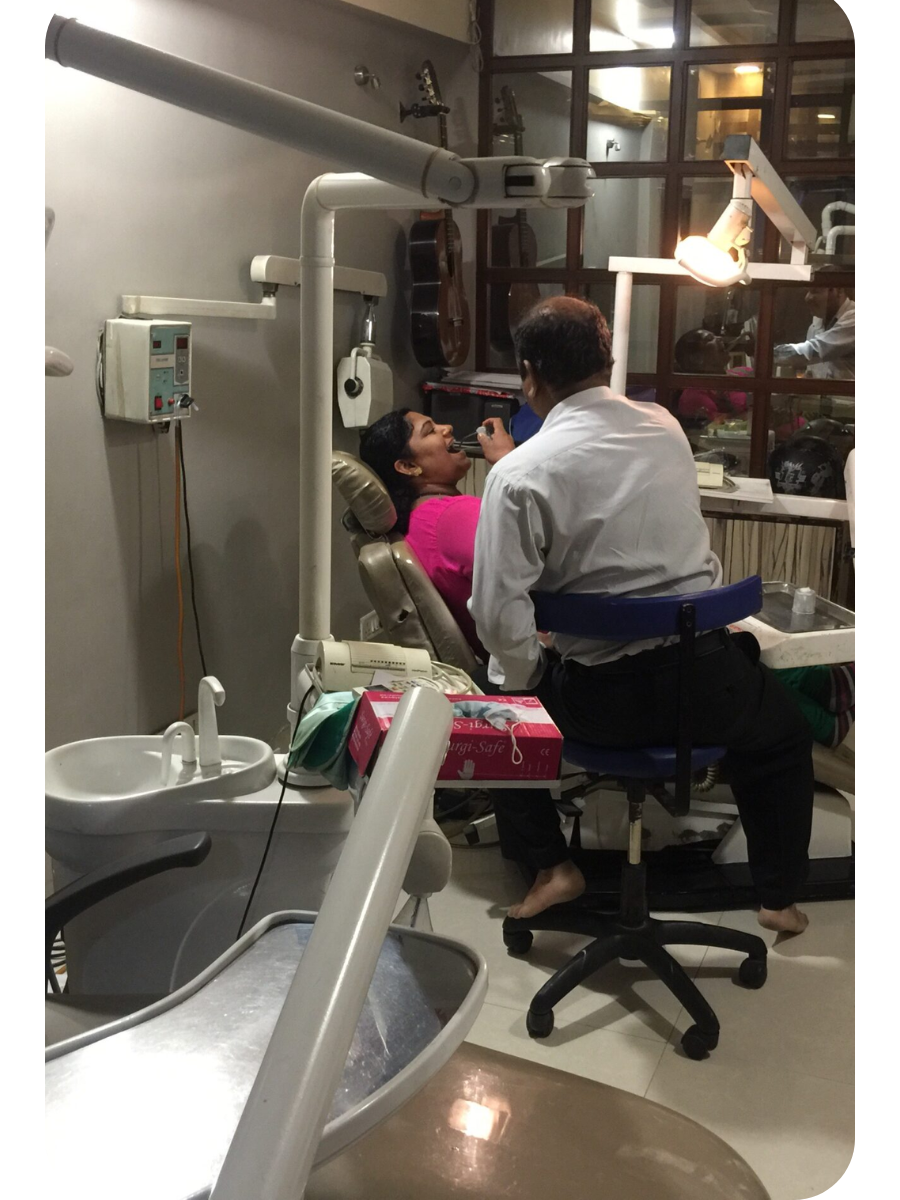
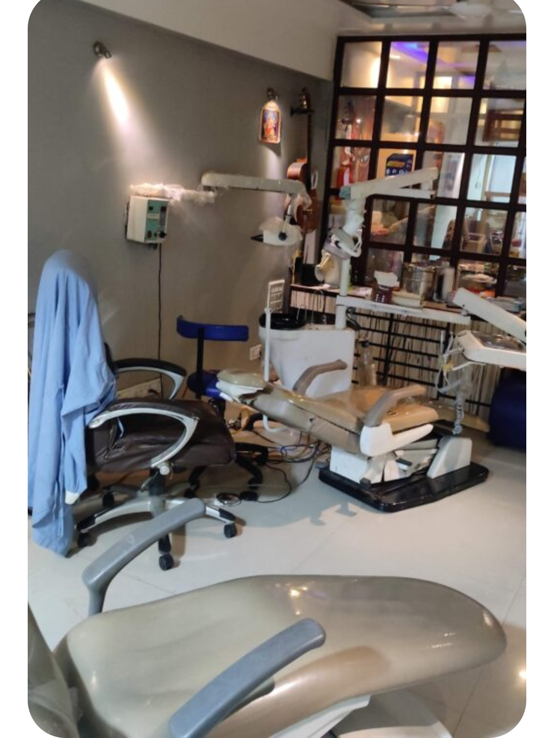
High End Equipments
We aim to deliver comprehensive dental care in a modern office setup equipped with high tech technology and equipment’s to ensure an unprecedented comfort to our patients.
WHAT WE DO
OUR PROCESS

What we do ?
WE CARE ABOUT YOU
Our clinic’s experts have extensive technical and academic knowledge in various areas of dentistry, ensuring that patients receive top-notch care and professional services. .

We give you
MEDICAL ADVICES
We believe in informed decision-making and provide thorough explanations of all dental procedures, including cosmetic, restorative, and preventative options, to empower our patients to make informed choices about their oral health.

We offer professional
MEDICAL SERVICES
We have the best experts completely ready to meet all of your needs for dental care right here in our clinic. We offer a range of dental care services from general family dentistry to root canal therapy, veneers, reconstructive and cosmetic treatments along with orthodontics services.
DOCTOR YOU CHOOSE
Decide what you are looking for in a doctor. A good first step is to make a list of qualities that matter to you.
REGULAR CHECK-UPS
We don’t forget to keep a check on our patients who are undergoing any treatment phase. It’s the most crucial part of our job to keep checking up on our patients.
MODERN FACILITIES
Our clinic is a full-service dental clinic that offers patients the best in quality dentalcare. Using state-of-the-art medical technology and proven methods of individualized care, our patients can be assured they will receive unsurpassed personal care and attention.
Working Hours
Monday
9:00 AM – 10:00 PM
9:00 AM – 10:00 PM
9:00 AM – 10:00 PM
9:00 AM – 10:00 PM
9:00 AM – 10:00 PM
9:00 AM – 10:00 PM
10:00 AM – 01:00 PM
PLAN A VISIT TO YOUR DOCTOR
We are always here to help you. Don’t hesitate and contact us to book your appointment.About Us
Best Dental Clinic That You Can Trust
We are a one-stop clinic equipped with state-of-the-art infrastructure to deliver specialized care to our patients with precision and perfection.
Complete Dental Care
Our panel of dentists are top rated MDS professionals with immense knowledge backed with experience. They are equipped to provide complete dental care under one roof.
Affordable Pricing
Our aim is to revolutionize dental care and provide top notch treatment to our patients in the most affordable and easily accessible way.
Call Today : +91 95949 00900/ 201,2nd floor,Accord Classic, Goregaon East, Mumbai, 400063
Open Hours Mon-Sat 8.00 am – 8.00 pm Sunday by appointment
It’s So Fast
Yoga Asanas for Oral Health | Improve Your Dental Health Today
Yoga Asanas for Oral Health Do you want to improve...
Read MoreFebruary 13, 2023“The Importance of a Properly Fitted Sports Mouth Guard: A Guide by Dr. Rajkumar, Dentist in Goregaon East Mumbai”
Introduction: As a dentist practicing in Goregaon East, Mumbai at...
Read MoreFebruary 9, 2023“The Evolution of Root Canal Irrigants in Dentistry: Understanding the Progress with Dr. Rajkumar Singh “
Introduction: Root canal treatment is a crucial procedure in dental...
Read MoreFebruary 8, 2023“Maximize Your Smile with Self-Ligating Braces at Baghel’s Dental World, Goregaon East, Mumbai”
Unlock a Brighter Smile with Self-Ligating Braces: The Future of...
Read MoreFebruary 8, 2023Brand We Use